For generations, medical students were taught that the map of the human body was essentially complete. Yet cancer imaging has now exposed a hidden structure in the head and neck that behaves like a distinct organ, forcing anatomists to redraw that map. The discovery, rooted in prostate cancer research, is reshaping how specialists think about radiation, surgery, and even how we count the organs that keep us alive.
What began as a technical advance in tumor scanning has turned into a fundamental shift in basic biology. By following an unexpected glow deep behind the nose and throat, researchers have identified a previously overlooked salivary gland system that appears to play a crucial role in lubricating the upper airway and protecting patients during treatment.
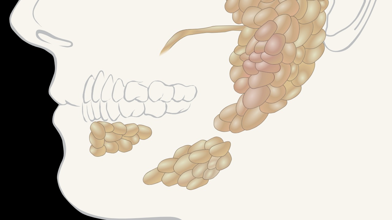
The hidden glands that lit up on cancer scans
The story starts with a tool designed for a very different purpose. While refining prostate cancer diagnostics, scientists at the Netherlands Cancer Institute began using PSMA PET/CT, a hybrid imaging technique that tags a prostate-specific membrane antigen and tracks it through the body. While scanning prostate cancer patients, they noticed two large, symmetric areas of activity in the upper throat that did not match any known structure, a pattern later echoed in a separate description that imaging had revealed a new organ hiding in the human head while using PSMA PET and CT scans at the Neth research center. Those bright spots, sitting where no textbook organ was supposed to exist, prompted a closer look at the anatomy behind the glow.
Follow up examinations showed that the mysterious structures were tucked in the back of the nasopharynx, near the torus tubarius, in a region that is notoriously difficult to visualize with conventional tools. Researchers at the Netherlands Cancer Institute reported that this previously unknown gland in the back of the nasopharynx appeared consistently in patients undergoing PSMA PET/CT, suggesting it was not a tumor or artifact but a normal part of human anatomy. The same pattern was highlighted in social media summaries of imaging that described how PSMA PET scans, typically used for prostate cancer, had unexpectedly outlined a new organ in the head, reinforcing that a cancer-focused technology had stumbled onto a basic anatomical blind spot.
From curiosity to “tubarial” salivary organ
Once the glow was recognized as real tissue, anatomists moved quickly to characterize it. Detailed dissection and histology showed that the structures contained mucous glands and ducts consistent with salivary tissue, but in a location that had not been cataloged as a major gland. In 2020, a formal description introduced the term “tubarial salivary gland” for this new pair of organs between the nasal cavity and throat, emphasizing that they formed a distinct salivary entity rather than a minor cluster of cells. An abstract on the tubarial salivary gland described it as a new kid on the block among salivary organs, underscoring that it sat alongside, not inside, the classic parotid, submandibular, and sublingual glands.
Independent commentary on the finding framed it as part of a broader realization that there might be two new organs hiding inside the head, both linked to salivary function and quality of life. Analysts noted that what we had long called “minor” glands in the nasopharynx were, in fact, organized into a coherent structure with its own ducts and physiological role, and that scientists were now debating how these tubarial glands fit into the traditional list of organs that make you enjoy life. A focused review of the tubarial salivary gland stressed that the latest salivary glands were identified through advanced imaging and then confirmed with targeted anatomical work, a sequence that turned a radiologic curiosity into a named organ system.
Why cancer doctors care about a new salivary organ
For oncologists, the discovery is not just a naming exercise, it is a practical warning sign. Radiotherapy to the head and neck often damages salivary tissue, leaving patients with chronic dry mouth, difficulty swallowing, and a higher risk of infections. When researchers mapped radiation fields against the newly described tubarial glands, they found that standard treatment plans frequently bathed this region in high doses, which could explain some of the stubborn side effects that persisted even when known glands were spared. Reporting on the unknown gland in the back of the nasopharynx noted that the team at the Netherlands Cancer Institute had uncovered the structure while studying radiotherapy and oncology, and that its location made it particularly vulnerable during cancer treatment.
The implications extend beyond a single hospital. A detailed account from the Netherlands Cancer Institute explained that cancer researchers had discovered a new salivary gland while evaluating PSMA PET/CT scans, and that recognizing this organ could help refine radiation planning to reduce complications. A separate research summary from a national biomedical database highlighted how the same PSMA PET/CT technique used in prostate cancer had unexpectedly illuminated salivary tissue in the nasopharynx, prompting calls to adjust dose constraints and contouring guidelines. In a popular science report, experts suggested that identifying this unknown human organ could help doctors better protect patients from treatment-related damage, especially in cases where head and neck tumors sit close to the newly mapped glands.
A pattern of “new” organs, from mesentery to interstitium
The tubarial glands are not the first time modern tools have forced medicine to rethink what counts as an organ. Earlier in the past decade, pathologists argued that the mesentery, a fold of tissue that connects the stomach to the intestines and holds the lower digestive system in place, should be classified as a single continuous organ rather than a fragmented structure. That reclassification was significant enough that a global record keeper listed the mesentery as the newest human organ and noted that its recognition brought the number of organs to 79. Around the same period, researchers using high resolution microscopy described the interstitium, a network of fluid filled spaces in connective tissue, as a potential new organ, with some reports suggesting it might be the body’s 80th organ and emphasizing that scientists claimed they had found a structure we never knew we had.
These debates show how much definitions matter. In coverage of the interstitium, scientists explained that what had long been dismissed as simple connective tissue actually formed a dynamic, shock absorbing system that might influence how cancer spreads. Commentators pointed out that if the interstitium were accepted as an organ, it would join the mesentery in expanding the official list of body parts, and that the tubarial salivary gland would then sit within a growing club of recently recognized organs. Together, these cases illustrate a pattern in which new imaging and staining techniques reveal that familiar regions of the body hide complex, organized systems that meet the criteria for organ status once someone looks closely enough.
What this means for future anatomy and treatment
The discovery of the tubarial salivary gland also hints at how many surprises might still be hiding in plain sight. A social media post from Sep described how, while scanning prostate cancer patients with PSMA PET and CT at the Netherlands Cancer Institute, researchers detected two large, symmetric structures in the nasopharynx that had gone unnoticed in standard anatomy. A related explainer from Apr noted that the protein used in these scans also lights up saliva glands, and that, interestingly, additional glowing tissue appeared in the upper throat during examinations, suggesting a role in the process of eating food and swallowing. These accounts underscore that the organ was not found through exploratory surgery but through routine cancer imaging that happened to use a tracer with an affinity for salivary tissue.
More from Morning Overview