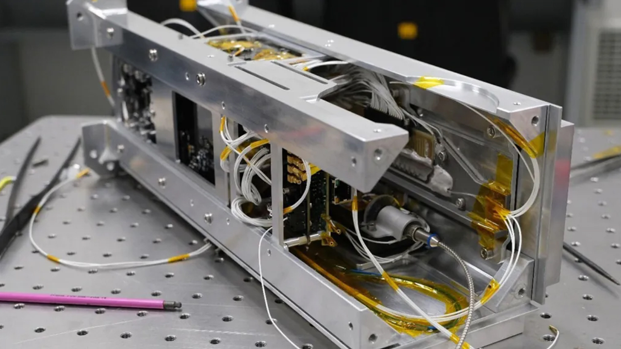
In a groundbreaking fusion of biology and technology, scientists have successfully 3D printed a tiny elephant inside a living cell. This remarkable feat not only showcases the potential of 3D printing at a microscopic level but also opens new avenues for advancements in medical science and cellular biology.
The Science Behind 3D Printing at a Cellular Level
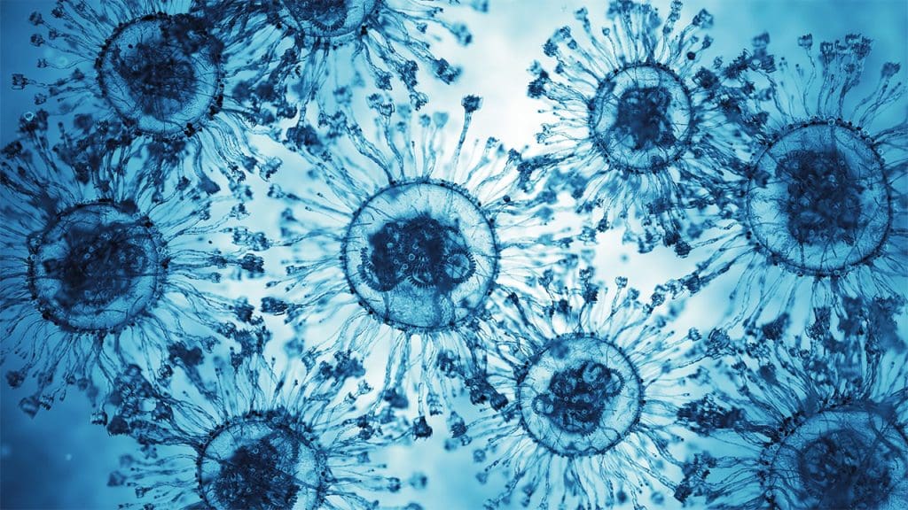
3D printing has transformed numerous industries by enabling the creation of complex structures with precision and efficiency. Traditionally, this technology involves layering materials to build objects from the ground up, based on digital models. However, applying this concept at a cellular level introduces unique challenges and opportunities. At its core, 3D printing within cells relies on adapting these principles to a microscopic environment, where precision and control are paramount.
To achieve this remarkable feat, scientists have turned to advanced techniques and materials that can operate on a nanoscale. The use of biocompatible materials is crucial to avoid damaging the delicate cellular structures. Additionally, the technology employs highly focused lasers to initiate the polymerization process, ensuring that the printed structures are not only incredibly small but also accurately positioned. Overcoming these technical hurdles has been key to making 3D printing inside living cells a reality.
The Experiment: Printing an Elephant Inside a Cell
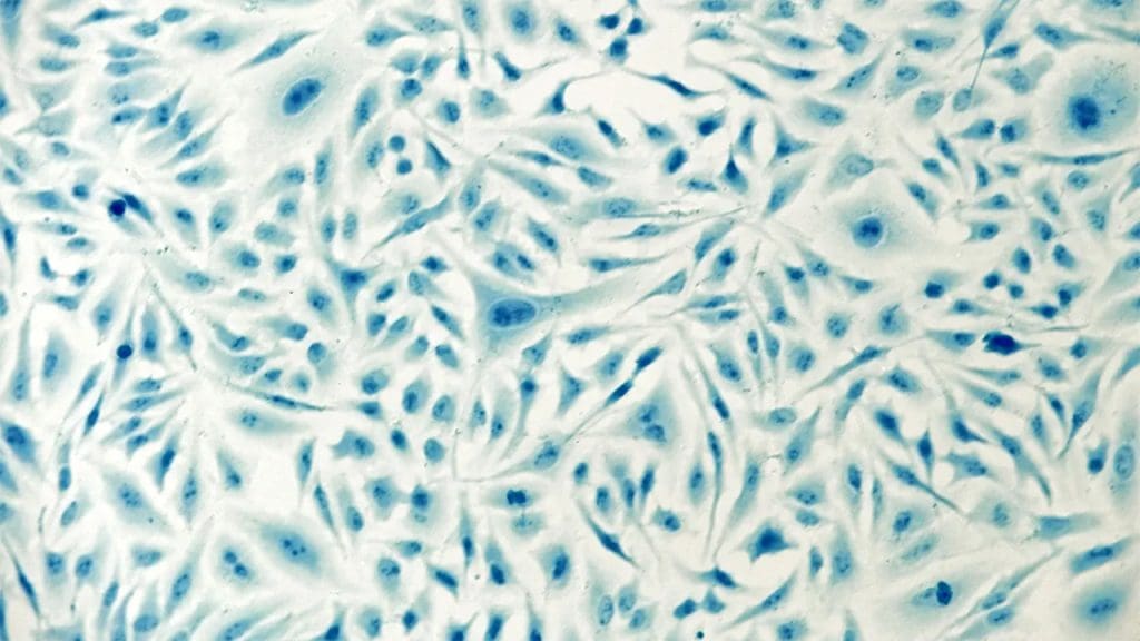
The experiment that resulted in a 3D-printed elephant inside a cell was a meticulously planned endeavor, highlighting both the sophistication of the technology and the creativity of the researchers. The setup involved suspending a single cell in a nutrient-rich environment to ensure its viability throughout the process. Using a highly advanced 3D printer equipped with a laser capable of etching at nanoscale resolutions, scientists were able to create the tiny elephant structure within the cellular matrix.
Choosing an elephant as the model for this experiment was both symbolic and strategic. The elephant, despite its large size in the natural world, served as a striking contrast to the minuscule scale of the cellular environment. This choice not only demonstrated the capability of 3D printing technology to replicate even the most unexpected shapes but also underscored the precision achievable at the microscopic level. The immediate outcomes of the experiment included the successful creation of the elephant model and valuable insights into the interaction between printed structures and their cellular hosts.
Implications for Cellular Biology and Medicine
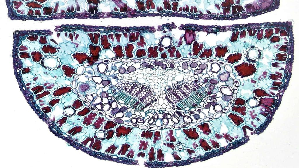
The ability to 3D print structures inside living cells opens a plethora of possibilities in the fields of cellular biology and medicine. One of the most promising applications lies in the development of drug delivery systems. By constructing microscopic delivery devices within cells, scientists could potentially direct pharmaceuticals precisely where they are needed, increasing efficacy while minimizing side effects. This development could revolutionize how treatments are administered, particularly for diseases requiring targeted intervention.
In the realm of regenerative medicine and tissue engineering, the technology offers new tools for constructing complex tissues and organs. Researchers could use 3D printing to create scaffolds that guide cellular growth, paving the way for the regeneration of damaged tissues or even the creation of entirely new organs. However, these advancements also bring ethical and safety considerations to the forefront. Manipulating cellular structures on such a fundamental level raises questions about long-term effects and the potential for unintended consequences, highlighting the need for thorough research and regulatory oversight.
Future Prospects and Research Directions
As 3D printing within living cells continues to evolve, the potential for future innovations is vast. Researchers are already speculating on the next steps, which could include increasing the complexity of the printed structures or integrating multiple types of materials to mimic more complex biological systems. These advancements could lead to breakthroughs in understanding cellular processes or even in developing new forms of life. The future of 3D bioprinting is undoubtedly bright, with each innovation building on the successes of previous experiments.
Key areas for further research include enhancing the resolution of printing technology, expanding the range of biocompatible materials, and improving the integration of printed structures with host cells. Interdisciplinary collaborations will be crucial, as the field requires expertise in biology, engineering, materials science, and more. By bringing together diverse perspectives and skill sets, scientists can push the boundaries of what is possible with 3D printing at the cellular level.
Public and Scientific Reactions

The scientific community has largely hailed the 3D printing of a tiny elephant inside a cell as a major milestone, sparking discussions about the future directions of nanotechnology and bioprinting. Researchers and experts from various fields have expressed excitement over the potential applications of this technology, recognizing its implications for advancing scientific understanding and medical practice. Conferences and publications have been abuzz with debates and papers exploring the myriad possibilities opened up by this breakthrough.
Public perception of this achievement has been equally noteworthy, with media coverage highlighting the whimsical nature of the experiment alongside its scientific significance. The idea of a tiny elephant inside a cell captures the imagination, drawing attention to the rapidly advancing capabilities of nanotechnology. This wider interest has the potential to inspire a new generation of scientists and innovators, eager to explore the intersections of technology and biology. As public awareness grows, so too does the support for continued research and investment in these cutting-edge technologies.