Neuroscientists have long dreamed of watching thoughts unfold as they happen, without disturbing the fragile tissue that produces them. That vision is suddenly closer to reality, as glowing neurons now let researchers follow brain activity from the inside out instead of relying on external flashes of light or bulky electrodes. The result is a new window into how individual cells and entire circuits work in real time, with implications that stretch from basic biology to future treatments for neurological disease.
At the center of this shift is a bioluminescent tool that makes brain cells light up on their own, turning the chemistry of neural firing into a visible signal. Rather than blasting the brain with lasers, scientists are teaching neurons to become their own lamps, revealing the ebb and flow of activity with unprecedented clarity and far less risk of damage.
From laser flashes to self-lit neurons
For years, the gold standard for visualizing brain activity has been fluorescent imaging, which depends on shining powerful light into tissue and reading the glow that comes back. That approach has yielded spectacular movies of neurons firing, but it comes with trade-offs: intense illumination can heat and stress cells, and scattering in dense tissue blurs the view. The new bioluminescent strategy flips the script by letting neurons generate light internally, so researchers can track activity without the same risk of phototoxicity or the need for invasive optical hardware.
In recent work, a team described a new bioluminescent tool that allows neurons to glow on their own, providing a direct readout of activity while avoiding the harmful illumination that has limited some fluorescent methods. By engineering brain cells to emit light only when key signaling molecules surge, the researchers can watch patterns of firing unfold in real time, even deep in tissue where external light would struggle to penetrate. The shift from externally driven fluorescence to self-generated bioluminescence marks a technical and conceptual break with past imaging, opening the door to longer, safer recordings of living brains.
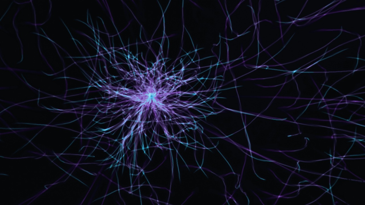
How CaBLAM turns calcium into light
The core of this advance is a molecular construct that translates the language of neurons into flashes of bioluminescence. When a neuron fires, calcium ions flood into the cell, triggering a cascade of events that underlie communication and plasticity. The new sensor, known as the Ca2+ BioLuminescent Activity Monitor, or CaBLAM, is designed to latch onto those calcium surges and convert them into visible light, effectively turning each spike of activity into a tiny beacon.
In a study published in Nature Methods, the team described CaBLAM as a bioluminescence tool that couples a calcium-sensitive domain to a light-emitting protein, producing a bright signal when calcium levels rise. Because the light is generated internally, there is no need for external excitation, which reduces background noise and allows cleaner detection of subtle changes in activity. The CaBLAM project is part of a broader push to understand how cells use calcium to regulate cellular activity, and it lives up to its name by turning those invisible ion flows into a readable optical code.
Watching single neurons behave in real time
One of the most striking aspects of this technology is its sensitivity at the level of individual cells. Traditional imaging often averages signals across many neurons, blurring the fine-grained dynamics that shape perception and behavior. By contrast, CaBLAM and related tools can capture the behavior of a single neuron as it responds to inputs, adapts to repeated stimuli, or participates in a larger network pattern, all without the need to insert electrodes directly into the cell.
Researchers report that the bioluminescent tool can capture the behavior of a single neuron with a bright, reliable signal that tracks calcium dynamics over time. Because the light output is tied tightly to the cell’s own activity, it becomes possible to follow how one neuron’s firing changes as an animal learns a task or experiences a new environment. That level of resolution is crucial for testing theories about how specific cell types contribute to memory, decision making, and mood, and it offers a way to link microscopic events to macroscopic behavior.
Glowing networks and the CaBLAM project
While single-cell recordings are powerful, the real promise of bioluminescent imaging lies in scaling up to entire networks. Brains compute through coordinated patterns of activity across thousands or millions of neurons, and any tool that hopes to explain cognition must eventually grapple with that complexity. By expressing CaBLAM across populations of cells, scientists can watch waves of calcium-driven light sweep through circuits, revealing how information moves and transforms as it passes from one region to another.
The CaBLAM project is explicitly framed as part of a larger effort to understand how cells use calcium to regulate cellular activity, and it is already being deployed in complex brain tissue. By mapping where and when neurons light up during specific tasks, researchers can infer which circuits are engaged and how their timing shapes outcomes. Over time, those glowing networks could help clarify why certain pathways are vulnerable in disorders such as epilepsy or neurodegeneration, and how targeted interventions might restore healthier patterns of activity.
Scientists make brain cells glow from within
At the heart of this story is a deceptively simple idea: instead of shining light on the brain, make the brain shine from within. Scientists have now done exactly that, engineering neurons so that their own biochemistry triggers light emission when they become active. The result is a kind of built-in reporter system, where each cell carries the machinery needed to announce its firing in real time, without external stimulation.
According to scientists who have made brain cells glow from within, this approach unlocks a safer and clearer way to watch the brain in action. By integrating the bioluminescent components directly into neurons, they can track activity in living tissue over extended periods, capturing both rapid spikes and slower shifts in baseline signaling. The work, which involves teams connected to the Carney Institute for Brain Science, underscores how genetic engineering and protein design are converging to turn neurons into self-reporting units, each one a tiny lamp revealing its own role in the larger circuit.
A new tool for watching neurons glow
Technically, CaBLAM is more than a clever acronym; it is a carefully tuned molecular machine. The construct combines a calcium-binding domain with a luciferase-like enzyme that emits light when it encounters its chemical substrate. When calcium levels rise during neural firing, the binding domain changes shape, activating the light-producing component and generating a burst of bioluminescence that can be detected with sensitive cameras. This design allows the sensor to respond quickly and reversibly, tracking the ebb and flow of activity with high temporal precision.
In their description of a New Tool for Watching Neurons Glow, the researchers emphasize that CaBLAM lives up to its name by delivering a bright, activity-dependent signal without the need for external illumination. That combination of sensitivity and gentleness is particularly important for long-term experiments, where repeated laser exposure could damage tissue or alter behavior. By relying on the cell’s own chemistry to drive light production, CaBLAM offers a more naturalistic way to monitor neural dynamics, one that is better suited to chronic studies of learning, development, and disease progression.
Linking bioluminescence to ultra-fast sensors
Bioluminescent tools like CaBLAM do not exist in isolation; they are part of a broader wave of technologies aimed at capturing the brain’s rapid, chemical conversations. Earlier this year, researchers combined ultra-fast sensors with advanced chemical imaging to track neurotransmitters moving between neurons in real time, effectively watching thoughts form at the level of synaptic communication. Those methods focus on the molecules that carry signals across gaps between cells, complementing calcium-based tools that monitor activity inside the cells themselves.By combining ultra-fast sensors with advanced chemical imaging, scientists have shown that it is possible to follow neurotransmitters as they cross synapses, breaking through a barrier that once made such measurements seem impossible. When paired with bioluminescent reporters that track calcium inside neurons, these approaches offer a more complete picture of neural communication, from the initial spike to the release and reception of chemical messengers. Together, they move the field closer to a unified, real-time map of how information flows through the brain during perception, thought, and action.
From single cells to whole-brain maps
As powerful as single-neuron imaging can be, the brain’s true complexity only emerges when activity is mapped across entire regions and, eventually, whole organs. That ambition has driven large-scale efforts to chart the structure and function of mammalian brains in three dimensions, creating reference atlases that can guide more targeted experiments. One landmark example is a comprehensive 3D brain map of the mouse, which required a massive, coordinated push across institutions and disciplines.
The work to build that map is described as the culmination of almost a decade of research by 150 scientists at 22 institutions led by the Allen Institute for Bra, in collaboration with Baylor College of Medicine and Princeton University. Those structural maps provide the scaffolding on which functional tools like CaBLAM can be overlaid, allowing researchers to place glowing neurons within a precise anatomical context. As bioluminescent imaging matures, it will likely be integrated into similar large-scale projects, helping to connect the dots between static wiring diagrams and the dynamic patterns of activity that give rise to behavior.
What glowing neurons could mean for medicine
The immediate impact of bioluminescent tools is on basic science, but the downstream implications for medicine are hard to ignore. Many neurological and psychiatric disorders involve subtle disruptions in how neurons fire and communicate, patterns that are difficult to detect with coarse measurements like scalp EEG or standard MRI. By revealing how specific cell types and circuits behave in real time, glowing neurons could help pinpoint the origins of conditions such as epilepsy, autism spectrum disorders, or depression, and guide the development of more precise interventions.
Because the new bioluminescent tool allows neurons to glow on their own without harmful illumination, as highlighted in the description of a new bioluminescent tool, it is particularly well suited to long-term studies of disease progression and treatment response. Researchers could, for example, follow how neural activity patterns change as an experimental drug takes effect, or how circuits reorganize after a stroke or traumatic brain injury. While clinical applications will require careful testing and adaptation, the basic principle is clear: the more precisely we can see the brain at work, the better our chances of designing therapies that restore healthy function rather than simply masking symptoms.
More from MorningOverview