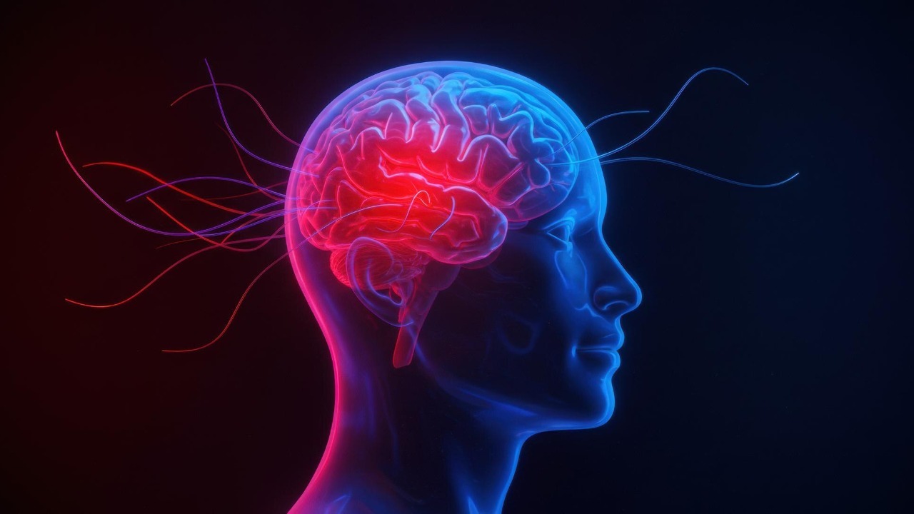
The human mind is turning out to be far stranger and more intricate than the tidy diagrams in old biology textbooks ever suggested. A wave of new research is revealing hidden layers in the brain’s memory center, unfamiliar electrical signals, and even memory-like processes in organs far from the skull. Together, these findings point to a startling secret: what we call “mind” is not a single thing in a single place, but a dynamic web of systems that stretch across the body and even challenge long-held assumptions about consciousness itself.
Instead of a static control room, the brain now looks more like a living city, with specialized districts for navigation, emotion, and awareness, all wired together by signals that scientists are only beginning to decode. As I trace these discoveries, from star-shaped cells that may store experience to kidney cells that behave like tiny archivists, a new picture emerges of how thought, memory, and identity might really work.
The brain’s hidden blueprint for memory
For decades, the hippocampus has been described as one of the brain’s major centers for memory, navigation, and emotion, but the internal wiring of this region was often treated as a black box. That picture is changing fast. Researchers have now identified a newly uncovered four-layer blueprint in the hippocampus that reorganizes how I think about memory storage and retrieval, showing that this structure is not a simple relay but a finely stratified hub where different layers appear to specialize in distinct aspects of experience, from spatial context to emotional tone, within one of the brain’s key memory centers.
This layered architecture does not sit in isolation. It fits into a broader surge of work on the mind and brain that is cataloging how memory, perception, and emotion are distributed across multiple circuits rather than locked into single “modules.” Top Headlines in Dec have highlighted how Scientists Find Hidden Layers in the Brain’s Memory Center, and those findings now sit alongside other reports of complex neural dynamics in the cortex and subcortex that are reshaping our understanding of cognition. When I scan the latest Mind & Brain News, I see Dec, Top Headlines, Scientists Find Hidden Layers, Brain, Memory Center, and Scientists all converging on the same message: the brain’s internal blueprint is richer and more layered than earlier models ever captured, as reflected in the evolving coverage of mind and brain.
The mind’s internal compass and our sense of self
One of the most striking advances in recent years is the discovery that the brain carries its own internal navigation system, a kind of built-in GPS that quietly tracks where we are and where we are heading. Earlier this year, Researchers reported that they had discovered two brain regions that act as a “neural compass,” keeping people oriented while they navigate a virtual city and helping them recognize various locations in the city, a finding that shows how spatial orientation is anchored in specific circuits rather than being a vague psychological feeling, as detailed in work where Researchers discovered this compass-like activity.
This internal compass is not just about finding a coffee shop in an unfamiliar neighborhood. It appears to be deeply tied to how we maintain a stable sense of self in a changing world. Studies on the brain’s “internal compass” show that when these orientation systems falter, people can experience disorientation and confusion that feel as if the world itself has slipped out of alignment. These discoveries illuminate the mechanisms by which the brain adapts to varying surroundings and explain why disruptions in these circuits can cause people to experience feelings of disorientation and confusion, sharpening our picture of the brain’s internal compass and how it stabilizes everyday experience, as shown in work on unlocking the internal compass.
Consciousness moves to the back of the brain
For a long time, popular accounts of neuroscience placed consciousness in the brain’s frontal lobes, treating the front of the brain as the executive suite where awareness supposedly lives. That view is now under direct challenge. According to IIT, a leading theoretical framework, the seat of consciousness is instead likely to be in the sensory representation in the back of the brain, and not in the frontal regions that are more involved in planning and control, a claim that has set up a high-stakes debate over how to interpret brain imaging and stimulation studies, as summarized in analyses that note how, According to IIT, the back of the brain may carry the richest content of experience, as described in work on consciousness ideas.
New experiments are backing up that shift in emphasis. In a recent “epic brain showdown,” researchers compared competing models of consciousness and found Surprising Clues From the Back of the Brain The study uncovered key functional connections between neurons in the brain’s posterior regions that appear to contribute heavily to the richness of what we see, and this challenges the long-standing idea that the front of the brain contains the full, detailed content of our visual experience, suggesting instead that sensory areas in the back of the brain may be doing more of the heavy lifting for awareness than previously thought, as highlighted in reports on Surprising Clues From the Back of the Brain The and further elaborated in coverage that notes how this work challenges the long-standing idea of a purely frontal seat of consciousness.
Signals that make human neurons stand out
At the microscopic level, scientists are discovering that human neurons do not simply fire like on-off switches. Brain cells, or neurons, link up through long, branching wires and shuttle messages along these cables to communicate with other neurons, but in human tissue researchers have now identified a unique kind of brain signal that appears to be absent in the brains of primates, our evolutionary cousins, suggesting that human neurons may process information in a more complex way than previously assumed, as described in work that details how, in Jan, Brain cells were found to carry a newfound signal in human neurons, as reported in studies of a newfound brain signal.
Other teams have gone further, showing that certain cells in the human brain transmit signals in a way not seen in corresponding rodent cells, a difference that emerged in a new study published in Science and that underscores how human neural computation may rely on mechanisms that standard animal models do not capture. In that work, the journal Science is cited as the venue where researchers reported that these human-specific signaling patterns could allow individual neurons to solve problems that scientists previously thought required entire networks, a finding that has prompted calls to rethink how we model cognition and disease in animals versus humans, as outlined in reports on how a new kind of signal was identified in the human brain and published in Science.
Star-shaped cells and the quiet architecture of memory
While neurons have long been the stars of neuroscience, a different kind of cell is now stepping into the spotlight. In a series of studies, scientists have focused on star-shaped brain cells known as astrocytes and found that these cells may underpin the brain’s massive memory storage, acting as a kind of scaffolding that supports and modulates neural networks rather than simply serving as passive support tissue. A new machine learning model shows that star-shaped brain cells may underpin the brain’s massive memory storage and could inspire advances in AI and Alzheimer’s research, suggesting that the brain’s memory capacity might depend as much on these glial networks as on the firing of neurons themselves, as described in analyses of how a Star-shaped brain cell model reshapes thinking about storage.
At MIT, researchers have pushed this idea into a bold new framework. An MIT breakthrough has proposed that star-shaped brain cells could be the secret behind human memory, with MIT scientists suggesting that these cells, with their intricate networks and calcium signaling, might encode and integrate information across wide swaths of cortex, potentially far more than neurons alone could. In this view, astrocytes are not just helpers but active participants in the encoding of experience, a shift that could transform how we design drugs for memory disorders and how we build artificial systems that mimic human learning, as detailed in coverage of an MIT breakthrough and in discussions where MIT scientists have proposed a bold new model of astrocyte-driven memory.
Memories that escape the brain
Perhaps the most unsettling twist in the new science of mind is the realization that memory-like processes are not confined to the nervous system at all. Scientists found memory’s molecular machinery at work in cells outside the nervous system, including kidney cells that can form memories of past signals, a discovery that suggests the basic biochemistry of remembering is a more general feature of life than anyone expected. In these experiments, Scientists showed that non-neural cells can adjust their responses based on prior exposure, effectively storing information about past events in molecular circuits, as reported in work showing that Scientists found memory’s molecular machinery in kidney cells.
Other research has gone further, arguing that memories are not only in the brain. A groundbreaking study has found that cells outside the brain may have memory functions, challenging the long-held belief that only neurons can store experience, and pointing instead to a “memory gene” similar to brain cells that appears to operate in other tissues. In Nov, Mind, Blowing Discovery, Scientists Discover That Memories Are Not Only, Brain, and related work have framed this as a Mind-Blowing Discovery: Scientists Discover That Memories Are Not Only in the Brain, suggesting that the body may carry multiple overlapping archives of experience that interact with, but are not reducible to, neural circuits, as detailed in reports on how Mind Blowing Discovery, Scientists Discover That Memories Are Not Only, Brain and in follow-up coverage that emphasizes how cells outside the brain may share a memory gene.
When emotion hijacks rational thought
Even as these discoveries expand the map of where memory and consciousness might live, they also help explain why our thinking can feel so fragile in the moment. The human brain has two frontal lobes, both of which are situated at the front of the brain, and these regions are crucial for rational reactions to threats, for weighing options, and for making decisions. Yet under sudden stress, the amygdala can trigger what psychologists call an “amygdala hijack,” a surge of emotional response that temporarily overrides the frontal lobes and disrupts the careful rationalization of situations, a pattern that helps explain why people sometimes say or do things in anger that they later regret, as described in detailed accounts of how a rational reaction to a threat depends on the frontal lobes, which are the areas where a person rationalizes situations and makes decisions, as outlined in discussions of amygdala hijack.
Scientists have even watched this kind of disruption unfold in real time. In one line of research, Scientists have seen what is happening in the brain at the moment we get startled and lose our train of thought, capturing how a sudden noise or unexpected event can scramble working memory and derail ongoing mental tasks. These observations show that the brain’s delicate balance between focused attention and environmental vigilance can be tipped in an instant, which is why a car horn, a dropped plate, or a buzzing smartphone can make someone forget what they were about to say, as documented in studies where Scientists have seen the neural signature of a lost train of thought.
Illness, education, and the fragile roots of mental life
The same circuits that support memory and consciousness can also become sites of vulnerability. Indeed, in recent years scientists have made many exciting discoveries about the function and dysfunction of the human brain, tracing how subtle changes in connectivity, chemistry, or inflammation can tip a person from healthy cognition into mental illness. These findings show that conditions like depression, schizophrenia, and anxiety are not abstract labels but concrete patterns of altered brain activity and structure, and they underscore how understanding the roots of mental illness requires careful study of both normal and disrupted neural function, as summarized in analyses that begin with the observation that, Indeed, scientists have made major strides in mapping the function and dysfunction of the human brain, as described in work on the roots of mental illness.
At the same time, educators and neuroscientists are beginning to translate these insights into practical strategies for learning. Utilizing advanced imaging techniques and rigorous neuroscientific research, a wealth of insights has emerged that enhances our understanding of cognition, memory formation, and information processing, and this body of work is now feeding into “neuroeducation” approaches that aim to align classroom practices with how the brain actually learns. By focusing on technique, such as how spaced repetition, multisensory input, and emotionally engaging material can shape neural dynamics, these efforts promise to make education more effective and more humane, as outlined in research that begins with the phrase Utilizing advanced imaging techniques and goes on to detail how such work informs our understanding of cognition and memory formation.
Cutting-edge tools that let us watch thoughts form
None of these insights would be possible without a revolution in the tools used to study the brain. One striking example is two-photon holographic microscopy, which allows scientists to monitor and manipulate neural activity in living animals with unprecedented precision. In a typical setup, Transcript Begin with an awake mouse positioned under a two-photon holographic microscope integrated with spatial light modulators and calcium indicators that bind to calcium ions, a configuration that lets researchers watch individual neurons light up as the animal perceives, decides, and moves, effectively turning the brain into a living movie of electrical and chemical activity, as described in protocols that start with the phrase Transcript Begin and go on to detail this imaging method.
On the human side, researchers are now detecting signals that were once thought impossible to measure. Recent work has identified first-of-its-kind dendritic signals in the human brain that allow individual neurons to handle complex computations, redefining our understanding of brain computation and suggesting that single cells may be capable of tasks previously attributed only to large networks. These dendritic signals allow individual neurons to solve problems that scientists previously thought required entire circuits, a finding that could reshape models of how thoughts and decisions emerge from neural tissue, as reported in coverage of how these dendritic signals were detected.
From lab to life: what these secrets mean for how we live
As the science grows more intricate, it also becomes more personal. When I read that Scientists have detected a first-of-its-kind signal in the human brain and that star-shaped cells may underpin the brain’s massive memory storage, I see not just technical breakthroughs but hints of future therapies for conditions like Alzheimer’s disease and Parkinson’s disease. While such heterogeneity is interesting in relation to the complex structure of the hippocampus and different roles of its subregions, it also shows up in disease models, where intranigral infusion of human-alpha synuclein oligomers induces cognitive impairment in rats associated with changes in neuronal firing and neuroinflammation in the anterior cingulate cortex, a region that is affected in PD-MCI patients [78, 79], as detailed in studies that note, While such heterogeneity is interesting in relation to the complex structure of the hippocampus, it also has clinical implications, as described in research on While such heterogeneity and its impact on cognition.
These findings also intersect with some of the most profound questions people ask about life and death. At the University of Virginia, Researchers explore phenomena such as near-death experiences, children who remember past lives, altered states of consciousness, and other unusual reports, not to prove or disprove any particular belief system, but to push the boundaries of what we know about how the mind and brain work. By treating these reports as data rather than dismissing them outright, these teams hope to refine our models of consciousness and to test whether current theories can account for the full range of human experience, as outlined in programs where Researchers explore phenomena that sit at the edge of mainstream neuroscience.
More from MorningOverview