
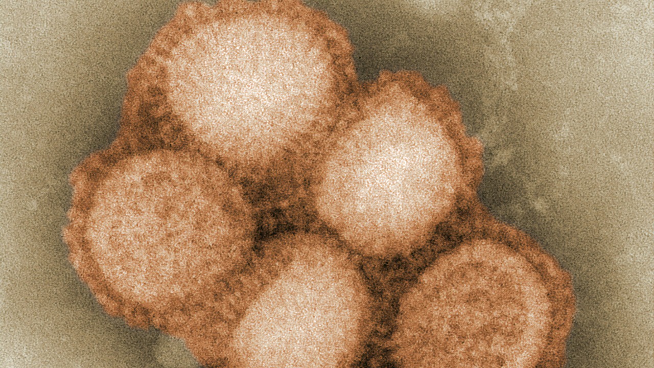
The first-ever image of PINK1 proteins represents a groundbreaking moment in Parkinson’s disease research. This pivotal achievement promises to enhance our understanding of the disease’s mechanisms, potentially paving the way for novel therapeutic approaches. By visualizing the PINK1 protein, researchers now have a clearer picture of how this enzyme functions within cells, offering insights that could lead to breakthroughs in treatment strategies.
The Role of PINK1 in Parkinson’s Disease
Overview of PINK1 Protein Structure and Function
PINK1, or PTEN-induced kinase 1, is a crucial protein involved in maintaining cellular health. Its primary role is to regulate mitochondrial quality control, ensuring cells function optimally. PINK1 achieves this by identifying damaged mitochondria and initiating their removal through a process called mitophagy. This is vital for energy production and overall cellular health, particularly in neurons, which are highly dependent on mitochondrial functions.
The structural visualization of PINK1 has revealed its complex architecture, which consists of distinct domains responsible for its kinase activity. Understanding these domains has been key to deciphering how PINK1 interacts with other cellular components. The new imaging provides unprecedented detail about the spatial arrangement of these domains, which could be crucial for designing drugs that can modulate PINK1 activity.
Genetic Mutations and Their Implications
Genetic mutations in the PINK1 gene have been closely linked to early-onset Parkinson’s disease. These mutations often lead to a loss of function in the protein, disrupting normal mitochondrial maintenance and contributing to neuronal death. The first image of PINK1 offers valuable insights into how specific mutations impact its structure and function. This understanding is critical for developing targeted therapies that can compensate for or correct these genetic defects.
Mutations such as the G309D and L347P have been extensively studied for their roles in altering PINK1’s functionality. With the new structural data, researchers can now visualize how these mutations affect the protein at an atomic level. This could aid in the design of small molecules or other therapeutic agents that specifically target these defective regions, offering hope for more effective treatments for those with hereditary Parkinson’s.
PINK1 and Mitochondrial Health
The connection between PINK1 and mitochondrial health is a cornerstone of Parkinson’s disease research. PINK1 acts as a sentinel, accumulating on the outer membrane of damaged mitochondria and marking them for degradation. This process prevents the accumulation of dysfunctional mitochondria, which can lead to cellular stress and neurodegeneration. The new image of PINK1 enhances our understanding of how this protein interacts with other mitochondrial proteins, such as Parkin, to facilitate mitophagy.
By elucidating the precise mechanisms of PINK1’s action, researchers can better understand its role in maintaining neuronal health. This knowledge is not only crucial for Parkinson’s disease but also for other neurodegenerative disorders where mitochondrial dysfunction is a key feature. The newfound structural insights into PINK1 could thus have far-reaching implications for understanding and treating a range of neurological conditions.
Technological Advances Leading to the Imaging Breakthrough
Cryo-Electron Microscopy (Cryo-EM)
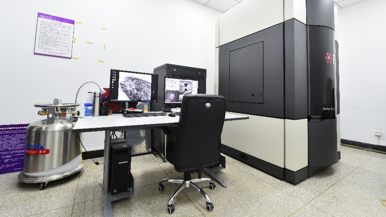
The visualization of PINK1 was made possible through advances in cryo-electron microscopy (Cryo-EM). This cutting-edge technology allows scientists to capture images of proteins at near-atomic resolution without the need for crystallization. Cryo-EM has revolutionized structural biology by enabling researchers to study proteins in a state that closely resembles their natural environment.
In the case of PINK1, Cryo-EM provided the clarity needed to discern its intricate structure and functional domains. The ability to capture such detailed images of PINK1 opens new avenues for drug discovery, as researchers can now design molecules that specifically interact with the protein’s active sites. This breakthrough underscores the transformative potential of Cryo-EM in understanding complex biological systems.
Challenges Overcome in Visualizing PINK1
Visualizing PINK1 was no small feat, as the protein is notoriously difficult to study due to its instability and tendency to aggregate. Researchers had to overcome significant technical and scientific challenges to stabilize the protein long enough to capture high-resolution images. Innovative approaches in protein purification and stabilization were critical in achieving this remarkable feat.
The interdisciplinary collaboration between structural biologists, biochemists, and computational scientists was key to overcoming these hurdles. By combining expertise from different fields, the team was able to develop novel methods for isolating and imaging PINK1, setting a new standard for protein research. This achievement not only provides a blueprint for studying other challenging proteins but also highlights the importance of collaborative efforts in advancing scientific knowledge.
Collaborative Efforts Across Disciplines
The successful imaging of PINK1 is a testament to the power of interdisciplinary collaboration. Researchers from various fields, including molecular biology, biophysics, and computational modeling, came together to tackle the complex problem of visualizing this elusive protein. By pooling their resources and expertise, they were able to achieve what was once thought impossible.
Such collaborative efforts are vital for scientific progress, as they bring together diverse perspectives and skill sets to address complex challenges. The PINK1 imaging breakthrough is a shining example of what can be accomplished when scientists from different disciplines work together towards a common goal. This achievement not only advances our understanding of Parkinson’s disease but also serves as a model for future research endeavors across the scientific community.
Implications for Parkinson’s Disease Treatment

Potential for Targeted Therapies
With the detailed structural information of PINK1 now available, researchers are poised to develop targeted therapies that can modulate its activity. Understanding the specific regions and mechanisms of PINK1 function allows for the design of drugs that can enhance or inhibit its activity, depending on the therapeutic need. This precision medicine approach could lead to more effective treatments with fewer side effects compared to current therapies.
For individuals with PINK1-related Parkinson’s, targeted therapies could correct the underlying genetic defects, potentially halting disease progression. The ability to precisely target the dysfunctional aspects of PINK1 opens up new possibilities for personalized medicine, where treatments are tailored to the specific genetic makeup of each patient. This marks a significant shift from the one-size-fits-all approach of traditional drug development.
Drug Discovery and Development
The imaging breakthrough is expected to accelerate drug discovery efforts aimed at mitigating or reversing Parkinson’s symptoms. By providing a detailed blueprint of PINK1, researchers can now screen for compounds that interact with the protein in desired ways. This structure-based drug design approach significantly speeds up the process of identifying promising drug candidates.
The availability of PINK1’s structural data also facilitates the use of computational modeling to predict how different compounds will interact with the protein. This can streamline the drug development process, reducing the time and cost associated with bringing new treatments to market. Ultimately, this breakthrough could lead to the development of more effective therapies that improve the quality of life for Parkinson’s patients.
Broader Impact on Neurodegenerative Research
The implications of the PINK1 imaging breakthrough extend beyond Parkinson’s disease, offering new insights into other neurodegenerative disorders. Many of these conditions share common pathways involving mitochondrial dysfunction, and the knowledge gained from studying PINK1 could inform research into diseases such as Alzheimer’s and Huntington’s.
By enhancing our understanding of mitochondrial biology, the PINK1 breakthrough could pave the way for novel therapeutic strategies across a range of neurodegenerative diseases. The ripple effects of this discovery underscore the interconnectedness of scientific research and its potential to drive innovation in unexpected areas. As researchers continue to build on this momentum, the future of neurodegenerative research looks increasingly promising.
Future Directions in Parkinson’s Research
Enhancing Imaging Techniques
As imaging technologies continue to advance, researchers are poised to gain even deeper insights into PINK1 and other related proteins. Future developments in Cryo-EM and other imaging modalities could provide even higher resolution images, revealing previously hidden details of protein structures. Such advancements will be crucial for understanding the intricate mechanisms underlying Parkinson’s and other diseases.
In addition to imaging, new techniques in protein engineering and synthetic biology could allow researchers to manipulate PINK1 and study its functions in greater detail. These approaches could lead to the discovery of novel therapeutic targets and strategies for treating Parkinson’s disease. The ongoing refinement of imaging and molecular techniques promises to keep pushing the boundaries of what is possible in biomedical research.
Expanding Genetic Research
The PINK1 imaging breakthrough opens up new avenues for genetic research aimed at uncovering additional links between the protein and Parkinson’s disease. By studying the genetic variations that influence PINK1 function, researchers can gain a deeper understanding of the disease’s underlying causes. This knowledge could inform the development of genetic tests for early detection and risk assessment.
Further genetic studies could also identify additional genes and pathways that interact with PINK1, providing a more comprehensive picture of the disease’s molecular landscape. This integrated approach to genetic research has the potential to uncover novel therapeutic targets and improve our ability to predict and prevent Parkinson’s disease. As researchers continue to explore the genetic underpinnings of the disease, the future of Parkinson’s research looks increasingly bright.
Building on the Current Momentum
The successful imaging of PINK1 has set the stage for a new era of Parkinson’s research, characterized by increased collaboration and innovation. This landmark discovery is likely to attract new funding and research initiatives, driving further advancements in the field. As more scientists join the effort to understand and treat Parkinson’s, the pace of discovery is expected to accelerate.
Building on the momentum generated by the PINK1 breakthrough, researchers are well-positioned to explore new therapeutic strategies and technologies. By harnessing the power of interdisciplinary collaboration and cutting-edge techniques, the scientific community is poised to make significant strides in the fight against Parkinson’s disease. The future holds great promise for patients and researchers alike, as they work together towards a common goal of finding a cure.